
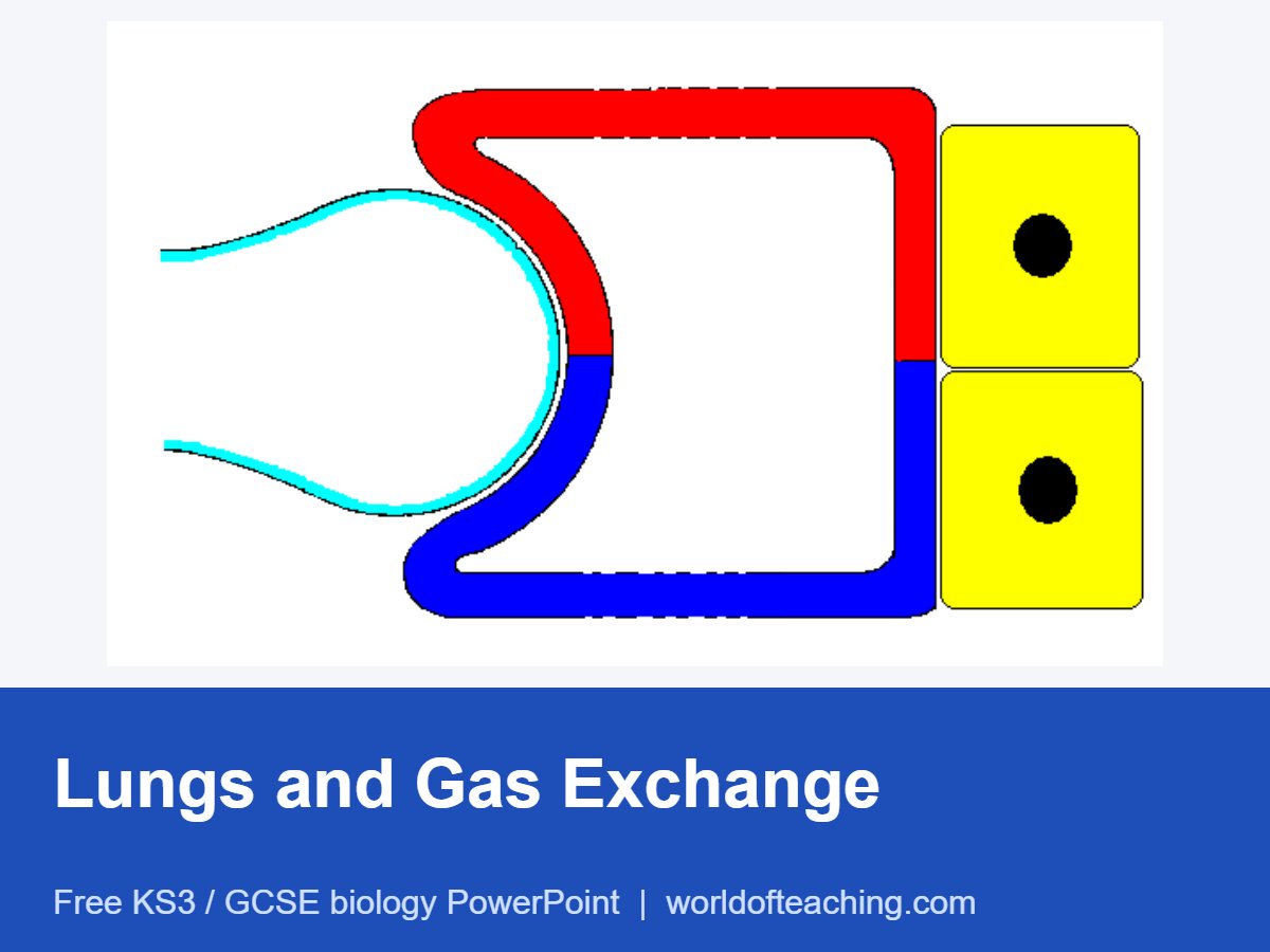
A lesson on the structure of the lungs and how gas exchange happens. It labels the trachea, bronchi and alveoli, then explains the features of alveoli and capillaries that make gas exchange efficient, with a recap of the learning objectives at the end.
What this presentation covers
- the structure of the lungs
- trachea and bronchi
- alveoli and their large surface area
- oxygen in and carbon dioxide out
- features of alveoli for gas exchange
- features of capillaries for gas exchange
Level: KS3 / GCSE
Slides: 9
Format: PowerPoint (.pptx)
Teaching notes
How to use it. A short 9 slide lesson that fits the gas exchange part of KS3 science and GCSE Biology (Organisation in AQA). It works well as a 20 minute introduction before a practical: a bell jar lung model shows how breathing works, and a sheep’s lung dissection or a video of one makes the size of the alveoli network memorable. Finish by asking pupils to explain how each feature of the alveoli speeds up diffusion.
Key words
- Trachea: the windpipe, held open by rings of cartilage
- Bronchi: the two tubes that branch off the trachea into each lung
- Alveoli: tiny air sacs where gas exchange takes place
- Gas exchange: oxygen diffusing into the blood and carbon dioxide diffusing out
- Diffusion: the net movement of particles from a higher to a lower concentration
Quick check questions
- Name the tiny air sacs where gas exchange happens.
- Give two features of alveoli that make gas exchange efficient.
- Why does carbon dioxide diffuse from the blood into the alveoli?
Show answers
- Alveoli.
- Any two of: a very large surface area, walls only one cell thick, a moist lining, and a rich blood supply.
- Its concentration is higher in the blood than in the air in the alveoli.
Related lessons: Blood | Aerobic and Anaerobic Respiration | Diffusion and Osmosis